GW Nanofabrication and Imaging Center
Bridging Life Sciences and Materials Sciences Research
The George Washington University (GW) Nanofabrication and Imaging Center (GWNIC) features state-of-the-art microscopy instrumentation and a newly-constructed Class 100 cleanroom. GWNIC provides university-wide core infrastructure for research in engineering, chemistry, physics, biology, public health, medicine and biomedical sciences. Located at the heart of GW’s Foggy Bottom Campus, the GWNIC is a catalyst for cross-disciplinary collaboration.
Nanofabrication
GWNIC features ~5,000 sq ft of Class 100 cleanroom with major instrument clusters for lithography, deposition, etching, measurement and characterization.
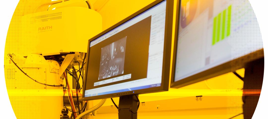
Imaging
GWNIC comprises two imaging suites (~5,500 sq ft) with the latest light, confocal and electron microscopes. Sample preparation equipment and services are also available.
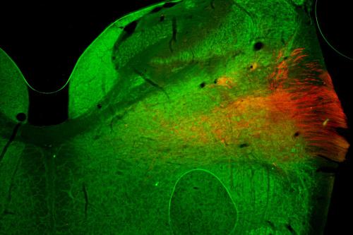
Featured Research
Researchers at the George Washington University discovered that a protein called Snail plays a key role in coordinating the response of brain cells after an injury. Read more in GW Today. Read the study, published in Proceedings of the National Academy of Sciences Nexus.
Segmentation performed on FIB-SEM images from corneas. Several individual axons are assigned different colors. One axon (aqua) moves in a different direction compared to the rest of the axons in the cluster shown. From Parlanti, P. et al., (2020).
Segmentation performed on FIB-SEM images from corneas. Intraepithelial Corneal Basal Nerves (ICBNs) are assigned the color yellow and the corneal epithelial basement membrane is assigned the color purple. From Parlanti, P. et al., (2020).
Image set of a spinal cord motoneuron produced by the Thermo Scientific Volumescope 2 Scanning Electron Microscope (SEM). Eleven of 350 sections were rendered using Imaris. Read more on LinkedIn.
A tiled and stitched volume of chicken vestibular ganglion, one hour after labeling with a fluorescent tracer. Image data was captured using a Leica SP8 FALCON and HC PLAN AP 20x/0.75 multi-immersion lens. Subsequent segmentation was performed with Imaris. Read more on LinkedIn.
A single hypoglossal neuron injected with biocytin during the course of patch clamp recordings. The imaging was performed on Leica SP8 Falcon. The dendrites were traced with Imaris. View the full paper. Read more on LinkedIn.
As demonstrated during our Correlative Microscopy Workshop in 2023, a workflow that utilizes the Thermo Fisher Teneo SEM and Maps 3 Software. Fluorescence image generated through z-stacking a large field of view using a Leica SP8 Falcon. The z-stack was then projected into a single image, inserted, and aligned using MAPS. Read more on LinkedIn.
A 3D volume of a mouse cornea using multiple stains, utilizing the Zeiss LSM 980 with Airyscan and Coherent Chameleon Discovery. The approach allows reliable tracing of small anatomical details. Read more on LinkedIn.