
Confocal Microscopy
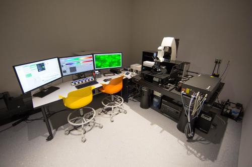
Carl Zeiss 710 Spectral Confocal Microscope

The Zeiss LSM 710 laser scanning confocal microscope can be used for long-term whole live-embryo, tissue slices and cell imaging at high
resolution. This upright microscope with x/y/z scanning stage, controlled via Zen software to execute complex multipoint temporal acquisition patterns, is supported by custom-designed on-stage incubation. This allows for long-term observations of explanted tissues and control of the temperature and environment either via perfusion or CO2 control.
- 32-channel spectral detector
- Two single channel photomultipliers and a photomultiplier on the forward pathway
- Six laser lines for excitation (458, 488, 514, 561 & 633 nm plus additional 405 nm diode laser)
- A customized high-end computer for large data sets (50GB).
- Objectives: W Plan-Apo 20x/1.0 DIC VIS-IR WD=1.8 water and 25x/0.8 multi-immersion LD LCI Plan-Apochromat (WD=0.55); Plan-Apochromat 10x/0.45 D=2.0; Plan-Apochromat 20x/0.8 M27 D=0.55; Plan- Apochromat 63x/1.40 Oil DIC; Alpha Plan Apochromat 100x/1.46
Carl Zeiss Cell Observer Spinning Disk Confocal Microscope

A Zeiss Cell Observer Spinning Disk Confocal microscope is fully integrated for live-imaging at high resolution. It allows for complex time-lapse
imaging experiments to be programmed and executed in high throughput manner.
- Two Photometrics Delta (64 fps, at 512x512) EM CCD cameras, for simultaneous acquisition of two channels
- Environmental control for CO2 and hypoxia
- A combination of high numerical aperture (i.e., 100x/1.46) and multi-immersion corrected objective lenses (i.e., 25x/0.8, 560 μ working distance)
- Virtual slides using high resolution/high magnification objectives (i.e., 100x/1.46 and 150x/1.35) and a color CCD camera
- Definitive focus
- Deconvolution, physiological analyses and stitching applications
Leica TCS SP8 Multiphoton Confocal Microscope
The Leica TCS SP8 Multiphoton Flexible Supply Unit features White LASER and 2-photon excitation. The White Light Laser is a fully tunable
supercontinuum laser with up to eight simultaneously usable lines in the range of 470 – 670 nm for maximal spectral flexibility in combination with AOBS and SP detector. Recording of two dimensional excitation and emission spectra turns the system into a spatially resolved fluorescence spectrophotometer for characterization. The White Light Laser offers pulsed excitation for FLIM with Pulsepicker for adjustable pulse intervals in FLIM.
This multiphoton microscope with high power femtosecond laser allows for deep tissue imaging. Precompensation for optimization of pulse width is fully integrated in LAS AF for easy control. It is equipped with Leica HyDRLD nondescanned detector. Its supersensitive photon detection with extraordinary quantum efficiency visualizes even faint details from deep tissue sections. The TCS SP8 SMD FLIM system combines the single photon counting technology FLIM with flexible confocal imaging. The system series integrates hardware and software from PicoQuant with the highend confocal system Leica TCS SP8 MP.
- Upright Stand DM6000 CFS flex
- Objective nosepiece H, 6pos,
- DM 6000 CFS stand Optical Outfit EL6000 with Transmitted Light Brightfield Detector
- SMD Software Package FLIM, 2 HyD SMD, FOV scanner SP8
- Switchable beam expander
- Scanhead: Two HyDs SMD, three Internal PMTs
- Laser 405 nm AOTF Flexible
- Laser Kit WLL2 + Pulse Picker
- IR Laser Chameleon Vision II, 680-1080nm