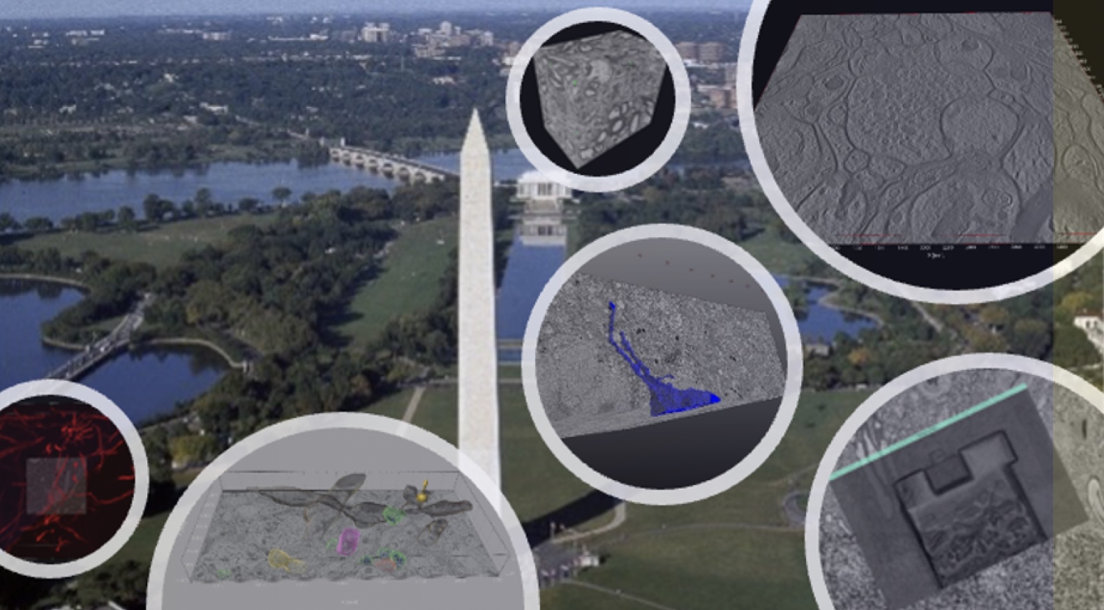
Workshop on Scalable Volume Electron Microscopy for Neuroscience

Overview
This workshop will focus on established and novel approaches for volume electron microscopy. The relationship between scalability and resolution will be the center of discussions. We will focus on solutions that enable the fusion of image data representing the global brain context linked to precise high-resolution volumetric sets. We will offer a comprehensive discussion on generating reliable volumes. Topics such as electron detection, noise, dosage, landing voltage, and sample depolymerization under the beam will be addressed, alongside the relationship between the physical section and electron-optical volume interaction.
We will also overview and provide practical demonstrations of four major approaches for volume electron microscopy – serial block face imaging with integrated microtome, FIB-SEM, array tomography, and axial tomography with STEM/TEM. Image analysis platforms, enabling segmentation and visualization will be discussed and demonstrated.
Leading experts from our industry partners will be presenting innovative approaches and will be available for discussions.
Highlighted demonstrations:
- Serial block face imaging using elastic scattered electrons
- Transmission Electron Microscopy/Scanning Transmission Electron Microscopy (STEM) Tomography
- Array tomography of post-embedding immunogold processed samples
- Sample preparation
- Image analysis
Additional Details
Monday, May 19 through Thursday, May 22; approximately 9:00 am-5:00 pm each day.
All four days of this workshop will be held on GW’s Foggy Bottom campus in the Science and Engineering Hall.
Registration
$800 registration fee.
Workshop Partners
The workshop is broadly supported by ThermoFisher Scientific, ZEISS microscopy, Johns Hopkins University Applied Physics Laboratories (BossDB), Leica Microsystems/ Thomas Scientific and DC Intellectual and Developmental Disabilities Research Center.
Questions?
Email gwnic gwu [dot] edu (gwnic[at]gwu[dot]edu) for more information.
Monday, May 19
| 8:00–9:00 am | Registration, coffee and light refreshments |
| 9:00–9:15 am | Welcome and brief logistics and safety introduction |
| 9:15 – 9:20 am | Overview of the workshop, agenda, etc. |
| 9:20–10:10 am | General Approaches for 3D electron microscopy. The neuroscience problem – size and resolution matter. Anastas Popratiloff, GWNIC |
| 10:10 – 10:15 am | Brief discussion |
| 10:15–11:00 am | Sample preparation strategies for volume EM. Strategies to link resolution and context. Cheryl Clarkson-Paredes, GWNIC |
| 11:00–11:15 am | Coffee break |
| 11:15 am–12:00 pm | Large-Scale Array Tomography for Brain Mapping and Beyond: Advanced Insights with Multi-Beam SEM. Joe Mowery, ZEISS Microscopy |
| 12:00– 1:00 pm | Lunch will be served and get together. |
| 1:00–1:45 pm | Volume electron microscopy with backscatter electron detection. Serial block face imaging with a microtome; focused ion beam imaging. Concepts, dosage, charging, z-resolution. Vivek Subramanian, Thermo Fisher Scientific |
| 1:45–1:50 pm | Discussion |
| 1:50–2:30 pm | Plasma-focused ion beam serial block face imaging Geoff Perumal, Thermo Fisher Scientific |
| 2:30– 2:45 pm | Discussion |
| 2:45–3:20 pm | 3D data logistics, from early postprocessing to robust 3D registrations. Approaches for the structuring of the image data in 3D. Data visualization and multivolume registration. Daniel J. Buss Amira, Thermo Fisher Scientific |
| 3:20–3:25 pm | Discussion |
| 3:25–4:00 pm | Local segmentation and machine training approaches. Chris Zugates Arivis, ZEISS Microscopy |
| 4:00–4:05 pm | Discussion |
| 4:05–4:20 pm | Coffee break |
| 4:20–5:00 pm | BossDB, The BRAIN repository for large-scale volumetric EM images. Segmentation and analysis on the cloud. Capabilities for secondary Analyses and community sharing. Erik C. Johnson Applied Physics Lab, Johns Hopkins University |
Tuesday, May 20
| 8:00–9:00 am | Coffee and refreshments |
| 9:00–10:30 am | “Hands-On” Critical steps in sample preparation.Keeping the brain architecture and orientation. Heavy metalen block staining procedure. Plunge freezing and freezesubstitution procedure. Sofia García-Hernández, GWNIC |
| 10:30–10:45 am | Coffee break |
| 10:45–11:30 am | Demonstration: Leica UC EnuityUltramicrotome. The power of automated serial sectioning. Mark Kukucka, Thomas Scientific |
| 11:30 am–12:15 pm | Seminar: Overview on the new technologies tooptimize large volumes reconstructions from serial sections |
| 12:15–1:15 pm | Lunch and social |
| 1:15–3:15 pm | Demonstration of block face imaging using Teneo FEG SEM/Volumescope – Sample installation and initial adjustments. Vivek Subramanian, Thermo Fisher Scientific |
| 3:15–3:30 pm | Coffee Break |
| 3:30–5:00 pm | Demonstration of block face imaging using Teneo FEG SEM/Volumescope – Sample installation and initial adjustments. Vivek Subramanian, Thermo Fisher Scientific |
Wednesday, May 21
| 8:00–9:00 am | Coffee and light breakfast |
| 9:00–9:30 am | Initial review of the volume scope data acquiredat night. Uploading to Amira software for final registration. |
| 9:30–10:45 am | Block face imaging with FIB-SEM – HeliosNanoLab 660 dual beam. Part 1: trench preparation andpositioning using large block face 2D image. Anil Kumar M.S. Thermo Fisher Scientific |
| 10:45–11:00 am | Coffee break |
| 11:00 am–12:15 pm | Setting up the serial block face imaging withSlice and View. The microscope will mill and image, and the process will be monitored in real-time. |
| 12:15–1:15 pm | Lunch break |
| 1:15–3:00 pm | Axial tomography with Talos 200 kV STEM.Preparation and scanning of 2D overview images to identifyareas of interest. Setting up parameters and running the scan. Sofia García-Hernández, GWNIC |
| 3:00–3:15 pm | Coffee break |
| 3:15–4:15 pm | Axial tomography post-processing and visualization |
| 4:15–5:15 pm | Plasma FIB demonstration from Nanoport in Oregon |
Thursday, May 22
| 8:00– 9:00 am | Coffee and light breakfast |
| 9:00– 10:00 am | Review of the FIB-SEM data, image registration using Amira, data visualization, and QC inspection |
| 10:00–11:30 am | Array tomography imaging using Helios NanoLab660 FEG SEM with immersion mode. Ultrathin sections with 10nm immunogold labeling of neurotransmitters. |
| 11:30–11:45 am | Coffee break |
| 11:45 am– 12:30 pm | BossDB, BRAIN initiative repository, and cloud image segmentation platform |
| 12:30– 1:30 pm | Lunch break |
| 1:30– 2:30 pm | 3D segmentation strategies using Amira. Overlay of different volumes (correlative visualization). 3D registration strategies. Lateral stitching, stitching artifacts, and imaging factors creating errors in 2D registration. Vertical (z) registration using direct processing and post processing with Amira. Trimming the volumes. Volume Visualization and inspection. |
| 2:30–3:15 pm | arivis Vision 4D for large volume rendering and segmentation |
| 3:15–4:00 pm | Coffee break |
| 4:00–5:00 pm | Strategies for rendering and visualization using Imaris |
| 5:00–6:00 pm | Closing reception with wine and cheese |
Workshop Location
All five days of the workshop will be held on GW's Foggy Bottom Campus in Science and Engineering Hall (800 22nd St NW, Washington, DC 20052).
Metro
Science and Engineering Hall is one block from the Foggy Bottom/GW DC Metro stop (Orange, Blue and Silver lines).
Maps and schedules are available on the Washington Metropolitan Area Transit Authority (WMATA) website.
Parking
Visitor parking is available in the Science and Engineering Hall Garage. The parking garage entrance is accessible via H Street between 22nd and 23rd Streets.
View more information about fees and other campus parking options.
Lodging
Local accommodations near the George Washington University
Follow the link above for a list of local accommodations near the George Washington University (GW). Special rates are available through the GW Hotels program. Link from this website to make your reservation or ask for the GW visitor rate when you register.
Options labeled "Foggy Bottom" will be closest to the workshop location.

